Reaction to study identifying neurons that restore locomotion after paralysis
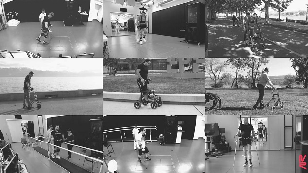
Research published in the journal Nature identifies the neurons that enabled nine paralysed patients to walk again after electrical stimulation of the spinal cord.
Juan de los Reyes - neuronas parapléjicos
Juan de los Reyes Aguilar
Head of the Experimental Neurophysiology Group at the Research Unit of the Hospital Nacional de Parapléjicos, member of the Castilla-La Mancha Health Service (SESCAM), the Castilla-La Mancha Health Research Institute (IDISCAM) and the Spanish Society of Neuroscience (SENC)
The study is of high scientific quality. It demonstrates the positive effects that epidural electrostimulation therapy has on patients with spinal cord injury. It also resorts to the use of animal models to reveal the cellular and physiological mechanism by which the expected effect is achieved.
In the article, the authors provide all the details, not only technical and experimental, but also, for example, the individual evolution of each patient treated, from his or her pre-therapy state to the final result obtained. This gives a true picture of the effectiveness of the therapy in the best cases and the possibility that in others (the fewest) it does not bring benefits.
In this sense, they show that in people with incomplete spinal cord injury (who preserve some connections between the spinal cord and the brain), the therapy makes it possible to recover walking through the application of epidural stimulation. In these patients, it is important to note that the continuous application of the therapy for 5 months manages to recover the voluntary action of walking without the need to apply the stimulation during walking (in 4 of 6 patients). These results must be considered a success.
However, people with complete spinal cord injury only managed to recover walking during the application of stimulation therapy. The difference between patients who regain function autonomously and those who can only walk when receiving epidural stimulation indicates the importance of preserving residual connections to brain structures and sensory inputs for optimal functional rehabilitation. Also the need to implement or combine other therapies to optimise outcomes in people with complete spinal cord injury.
Based on the results obtained in people who recover the ability to walk voluntarily, without the aid of epidural stimulation, the authors ask what happens in the spinal cord to recover this function in such an impressive way. To answer this question, they study the level of neuronal activity within the spinal cord using magnetic resonance imaging. As a result, they observed that in people receiving epidural stimulation, neuronal activity in the spinal cord is lower. This result is quite counter-intuitive, since a recovery of function should imply a higher neuronal activation. Based on these results, we propose to decipher the neuronal activity in the spinal cord in the situation of spinal cord injury with and without epidural stimulation treatment.
It should be noted that, in humans, experiments to identify neuronal types cannot be performed. Therefore, the authors use a model of spinal cord injury similar to that suffered by humans, but applied to mice. In these experimental animals, access to neuronal tissue is available for detailed study and for manipulation of neuronal activity using genetic techniques. The authors use the latest technologies in molecular biology and bioinformatics to characterise the molecular and spatial profile of RNA expression of all the cell types that make up the spinal cord under normal conditions and after spinal cord damage. An artificial intelligence computer procedure, developed by the authors and called "Augur", is applied to these data to identify whether a cell type responds with increased RNA production to a stimulus that alters cell physiology. The combination of both approaches, molecular biology and bioinformatics, allows the identification of the region of intermediate laminae in the spinal cord as the location of the neurons of interest, with the neuronal type identified as SCVsx2::Hoxa10. From the identification of the neuronal type that responds to therapy, the activity of these neurons can be manipulated by using optogenetic and pharmacogenetic techniques, which allow the target cells to be selectively activated or deactivated during the application of therapy and without the application of therapy. This confirms that this cell type is key in the process of gait recovery in response to epidural electrostimulation in animal models and in humans.
The relevance for the future is that, when the cell type that produces an effect is known, different therapies (single or combined) can be targeted to manipulate cell activity and achieve better results.
The authors are cautious because, knowing the complexity of cell types in the spinal cord, they consider that this is only one of the types involved in the process, and there could be other groups of neurons involved in different aspects of recovery. They also acknowledge that the work has been done in animals, and it is possible that different cell types exist between different species. Therefore, it will be necessary to confirm later (in postmortem tissue in humans) that these cells are modified by the effect of epidural therapy.
The study has obvious limitations, for example, recovery of function independent of stimulation only occurs in people with incomplete injury (4 out of 6 people). Whereas in people with complete injury (3 people), recovery of function is only observed during the time of therapy. Therefore, the efficiency of the therapy applied depends on the degree of preservation of nerve pathways connecting the spinal cord and brain. This highlights the importance of assessing the preserved post-injury spinal cord function in each individual in order to apply a specific or personalised treatment. Furthermore, although the efficiency of the therapy applied has been demonstrated, it is true that the patients become fatigued during the execution of the gait, covering a still reduced distance. One of the ways to improve the effects and duration of the therapy would be to combine it with other tools or pharmacological treatments, but currently access to the spinal cord by different routes for combined therapy is not developed.
However, the prospects for better results are very good and possible. The steps that Dr. Courtine's group has taken in this field have been solid and this work is an important advance built on a large knowledge base, with the prospect of optimising rehabilitation therapy for people with spinal cord injury. It should be considered from here that optimisation of therapies can be achieved by combining epidural stimulation with better implants and appropriate stimulation protocols to generate longer lasting effects. It also opens up the possibility of applying combined treatments, e.g. epidural stimulation together with pharmacological treatment targeting different cell types. Finally, taking into account that each person has a different spinal cord injury and preserves different degree and types of connections to the brain and sensory inputs, the prospect of developing personalised medicine for the treatment of spinal cord injury will be the next step.
- Research article
- Peer reviewed
- People
- Animals
