Reactions to study revealing how oocytes survive toxic protein aggregates for decades
An international team, led by a Spanish group, has published the mechanism that allows immature egg reserves (oocytes) to survive for many years, up to almost half a century in the case of humans. The research studies how oocytes are affected by protein aggregates similar to those that damage other cells such as neurons and can cause neurodegenerative diseases such as Alzheimer's disease. The finding of how these egg reserves are kept healthy may help to understand some causes of infertility. The results are published in the journal Cell.
Rocío Núñez Calonge - ovocitos proteínas tóxicas EN
Rocío Núñez Calonge
Scientific Director of the UR International Group and Coordinator of the Ethics Group of the Spanish Fertility Society
Oocytes, along with neurons and their extensions (axons), are the largest cells in the human body, and have in common that they do not divide, remaining in the body for many years.
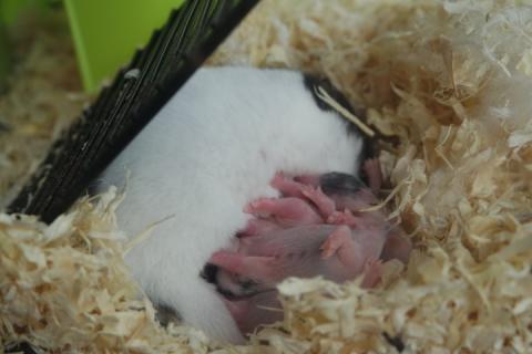
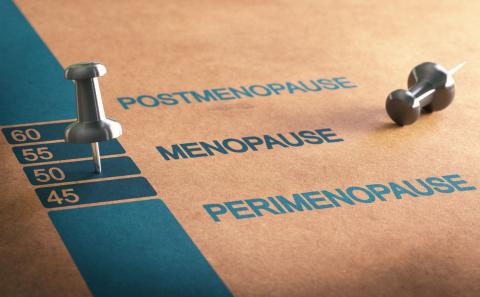
Oocytes, the female reproductive cells, begin to form in the ovary of the female foetus, and do not complete their maturation until puberty is reached and they are ready for fertilisation. In the newborn girl, there are almost a million oocytes, which degrade until the first menstruation or menarche. Of the nearly half a million oocytes in the adult woman, only a few will mature and become ready for fertilisation during her lifetime: one for each monthly ovulation. This process gives an idea of the fragility of these cells, which are indispensable for the perpetuation of the human species. Until now, little was known about the mechanism by which oocytes manage to remain intact for so many years: until the woman's menopause.
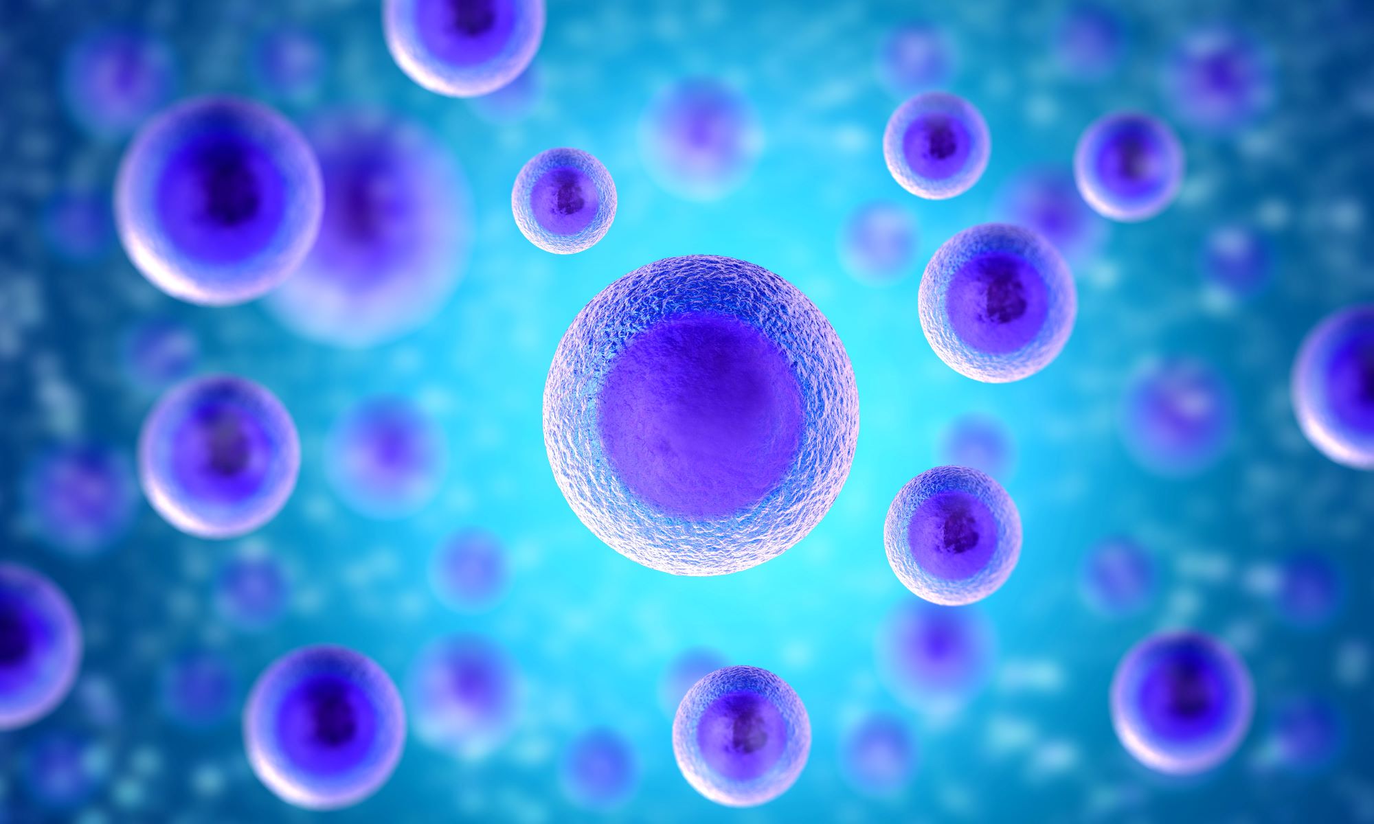
Oocytes consist of a nucleus, which contains the genetic information that will be passed on to offspring, and a cytoplasm, which serves as the cell's nutrient and energy machinery. The cytoplasm contains protein aggregates, in some cases harmful to the cell.
Long-lived, non-dividing cells, such as oocytes and neurons, are particularly sensitive to this accumulation of protein aggregates, as these cells cannot dissipate the aggregates by cell division. Although the mechanisms of formation, accumulation and physiological removal of protein aggregates in neurons have been studied in depth, it is not known how mammalian oocytes cope with this problem.
Zaffagnini et al. have published a study with mouse oocytes in which they have found how protein degradation occurs. The study is very good, very well documented both experimentally and in previous studies, and with very clear pictures.
Mouse oocytes store protein aggregates in compartments called endolysosomal vesicle assemblies (ELVA), where the aggregates are degraded during oocyte maturation. Functional assays revealed that, in immature oocytes, ELVAs sequester aggregated proteins and degrade them during oocyte maturation. Inhibition of ELVA degradative activity leads to accumulation of protein aggregates in the embryo and is detrimental to its survival. Therefore, ELVA represents a strategy to safeguard the functionality of these cells.
Poor oocyte quality is one of the main causes of female infertility. In many cases the poor quality is due to genetic defects associated with age, but there are other unknown factors that alter their viability and survival. Therefore, this research opens an interesting avenue for future studies to explore whether protein degradation and its misregulation could explain the age-related deterioration of oocyte quality.
However, there are still many questions, which the authors themselves raise: Could ELVA-like compartments exist in other cell types? It has been postulated that a similar mechanism could occur in neurons. But how these long-lived cells, oocytes and neurons, transport their protein aggregates to specialised compartments and regulate their degradation, and whether this actually happens in neurons, remain future avenues of research to be explored.
The main limitation is that this has been studied in mouse oocytes and it remains to be seen whether this is indeed the case in humans. The article compares it with neurons because they share the similarity with oocytes in that they do not divide and are long-lived, but the aim of the study and the focus is on oocytes and their future application in cases of female infertility. I would not, as in the case of the press release, talk about Alzheimer's disease. In fact, the article does not even mention it.
Antonio Urries - ovocitos proteínas tóxicas EN
Antonio Urries
Director of the Assisted Reproduction Unit at the Quirónsalud Hospital in Zaragoza and member of the Quirónsalud Scientific Committee
Among the fertility problems we face in assisted reproduction units, there are 20% of cases in which no apparent reason can be found, which is why we call it sterility of unknown origin. These cases frequently cause problems in achieving pregnancy even when we use techniques such as in vitro fertilisation and transfer embryos with the correct genetic load into the mother's uterus.
This is due to the fact that the genetic load of the embryo is as important as its cytoplasm and we must take into account that the embryo shares the cytoplasm of the oocyte, especially during the first days of development.
From a reproductive point of view, this article is very interesting as it demonstrates how the presence of certain protein aggregates in the cytoplasm of the oocytes can cause poor oocyte quality and embryos with a low probability of becoming pregnant, even if they have a correct genetic load.
These protein aggregates have a toxic effect, widely studied in this and other studies, which the egg may have difficulty in eliminating as they are generally eliminated during the cell divisions that the egg does not begin to carry out until it is fertilised.
The novelty of this article lies in the discovery by the authors of special structures, which they have named EndoLysosomal Vesicular Assemblies (ELVA), dispersed in the cytoplasm of the oocyte in a number of approximately 50, capable of localising and inactivating these protein aggregates, rendering them harmless.
This inactivation occurs just as the egg completes its maturation process prior to fertilisation by the sperm. ELVAs would act as a cleaning team, eliminating these deleterious effects on the egg, keeping its cytoplasm in optimal conditions for successful fertilisation and subsequent development.
According to the study, the demonstration lies in the fact that if ELVA activity is inhibited, an accumulation of these protein aggregates in the egg cell occurs, damaging the survival and future viability of the embryo.
On the other hand, it has also been seen how these aggregates can have harmful effects on other long-lived cells with few cell division processes, such as neurons, and could be related to neurodegenerative diseases such as Alzheimer's, so the study of how ELVA behave in this type of cell could help in the research and treatment of other diseases.
Zaffagnini et al.
- Research article
- Peer reviewed
- Animals