Human brain organoids form neuronal networks in mice
A US-based team with Spanish participation has successfully transplanted cortical organoids—self-organizing fragments of lab-grown human brain tissue—into modified mice lacking most of their cerebral cortex, the brain layer responsible for functions ranging from cognition and language to attention and decision-making. The research, published in the journal Nature, shows that the human tissue survived, grew, and developed functional connections with the mice's brains and, extending beyond that, with the spinal cord. According to the authors, this could help generate new models for studying disease-related alterations in human brain circuits.
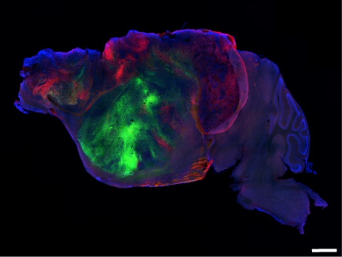
Side view of a xenocortical mouse brain showing nerve fibers extending from the human graft (colored with green and red fluorescent proteins) through the mouse brain (blue). Scale bar = 1 mm. Credit: S. Pasca lab, Stanford University.
Vega - Organoides cerebro
Iván Fernández Vega
Full professor of Pathological Anatomy at the University of Oviedo, Scientific Director of the Principality of Asturias Biobank (BioPA) and Coordinator of the Organoid hub of the ISCIII Biomodels and Biobanks platform
Does the press release accurately reflect the study?
"Broadly speaking, yes. The press release correctly conveys the study's key innovation: the creation of a model in which human cortical organoids grow and extensively integrate into the brain of a mouse that has undergone significant depletion of its own cortex. The graft comes to represent approximately 92% of the measured cortical tissue and establishes connections with various structures of the nervous system."
Is the study of high quality? Are the conclusions supported by robust data?
"Yes. It is a technically comprehensive study that combines histology and immunohistochemistry, single-cell and spatial transcriptomics, MRI, neuronal tracing, calcium imaging, electrophysiology, and behavioral studies. The use of independent approaches that converge on the same conclusion greatly strengthens the findings; the phenomenon is observed from multiple levels. Furthermore, the authors employ appropriate controls, analyze sex-based differences where possible, and note that experiments and analyses were conducted in a blinded manner whenever feasible.
Some of the most sophisticated experiments involve a small number of animals, so certain findings will need to be replicated in larger cohorts and with a greater number of cell lines."
How does this work fit with existing evidence?
"It represents a logical progression of the group's previous work. It had already been demonstrated that human cortical organoids transplanted into rodent brains could become vascularized, mature, and integrate better than in culture. The innovation here lies in eliminating much of the competition from the recipient animal's cortex, thereby creating a space where human tissue can grow much more extensively and form a widespread network. The goal, therefore, is not to replace conventional organoids but to overcome some of their major in vitro limitations."
Have confounding factors been taken into account? Are there significant limitations?
"The authors have made a considerable effort to control for confounding factors through the use of control groups, comparisons with non-grafted apallial mice, sex-based analysis, and various experimental approaches. However, significant limitations remain. The graft remains immature: it lacks canonical cortical lamination, complete arealization, and an interneuronal composition equivalent to that of the mature human cortex. Furthermore, although anatomical integration and neuronal activity are present, it cannot yet be claimed that the human neurons are necessary or sufficient to drive specific animal behaviors.
I would add another point from a pathological perspective: the graft increases in volume approximately 4.7-fold between the two- and three-month marks, yet the study was not designed to determine when this growth stabilizes. For models derived from pluripotent cells intended for long-term maintenance, characterizing proliferation kinetics and long-term safety will be important.
It is also interesting from the perspective of the 3Rs: here, the organoids do not replace the animal but rather integrate into it to create a human-animal hybrid model. This could help reduce animal usage if it allows for more data to be gathered per experiment, though that remains to be demonstrated."
What are the real-world implications?
"The immediate application is not clinical; this is fundamentally a new experimental model. Its primary utility could lie in studying neurodevelopmental processes, injuries such as perinatal hypoxia, and specific neurological diseases using human cells integrated into a living nervous system, followed by the evaluation of potential treatments. As a proof of concept, the study demonstrates that a hypoxic insult elicits a marked response in the grafted human tissue and measurable functional changes in the animal.
In the medium term, the value will lie in determining which human diseases the model truly recapitulates better than current models, and whether the results are reproducible across different iPSC lines, laboratories, and protocols."
Conflicts of interest: "I have no conflicts of interest related to this study. My scientific work involves organoid models, and I am a co-leader of the Organoid Hub at the Carlos III Health Institute."
Tim Viney - organoides cerebro
Tim Viney
This work builds upon their earlier study whereby they transplanted human cortical organoid tissue into the developing rat cerebral cortex demonstrating that the cells from the organoid integrated into the host brain.
“The advance here is they now use a genetic strategy to generate mice that have a depleted cerebral cortex, then transplant the human cortical organoid tissue into the brains of these mice. This remains technically challenging in terms of reducing variability when injecting the cortical organoid cells (4 injections per mouse), but they use fairly standard neurosurgical techniques that will be familiar to most rodent neurophysiologists/neuroanatomists.
‘Acortical’ mice are not a new concept, generated mice that lack a large proportion of the cerebral cortex. This remains an active area of research, e.g. Zheng et al used such mice to show they can still perform complex behavioural tasks, which makes for an interesting discussion on the roles of the cortex - the cortex only functions because it is integrated with the rest of the brain.
“I remain cautious about such studies, as these are high risk models that depend on the successful transplantation of viable cells that developed within the organoids. It is still not fully clear to me the methods for preparing the organoids for transplantation, but I think the cells from the organoids are not organised into ‘circuits’ when they are injected in to the mice, rather they are free in solution, hence it is tricky to understand how they become integrated in the host.
Note that the organoids themselves do not represent the complexities of the human brain, despite the expression of familiar molecular markers and structural characteristics of brain cells. The advantage of organoids comes from the genetic background of the donor, and the possibility to manipulate certain genes to see how mutations can affect particular cellular pathways. But they are not ‘brains in a dish’, as the connectivity is highly artificial (and typically simplified with minimal cell types). The authors themselves state as well the mismatch between the relatively rapid development of the mouse versus the much longer developmental trajectory of human cells.
They used a clever strategy to virally label cells in the organoids prior to transplantation, followed by adapting an existing technology of rabies tracing from the organoid graft. Otherwise, most of the techniques alone are quite standard, including behavioural tests, electrophysiology, histology.
The most informative and still gold standard way to investigate the human brain network organisation and activity is using acute ex vivo slice recordings - tissue is removed during neurosurgery then the living tissue can be kept alive (with all its local circuits intact) for at least 12 hours for physiological and anatomical studies. The other complementary technique is in vivo recordings in patients (e.g. epilepsy patients awaiting surgery).
Conflicts of interest: "I have no interests to declare except that I know one of the authors (Soltesz) who I regard highly. I have not worked directly with organoids but these are my views as a neuroanatomist/neurophysiologist."
Kaganovsky et al.
- Research article
- Peer reviewed
- Experimental study