Reaction: artificial intelligence outperforms imaging technicians in assessing cardiac function
The functioning of the heart can be studied by the percentage of blood it pumps with each beat, deduced by imaging techniques. Based on analyses by cardiologists, a US clinical trial concludes that an artificial intelligence model outperforms examinations initially performed by imaging technicians in terms of accuracy. According to the authors, the tool could "save physicians time and minimise the most tedious parts of the process".

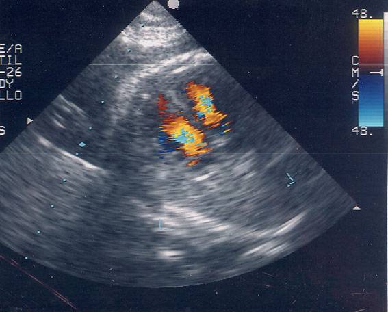
Ultrasound scan of a foetal heart. Photo: Hospital La Paz in Madrid
Pérez - IA (EN)
Julián Pérez-Villacastín
Head of Cardiology at San Carlos Clinical Hospital and professor at Complutense University of Madrid
The study is cleverly designed and I think it is a valid way to analyse what they are proposing. It is certainly one more piece of information that supports the use of new technologies as an aid in medicine. It is not at all surprising that through these technologies we are able to be more accurate, saving time and variability. In short, it is possible to be much more efficient thanks to these kinds of technologies.
In general, with everything related to imaging in the coming years we are going to see how this type of development surpasses human beings.
In Spain there are also centres where imaging technicians predominantly perform echocardiograms to assess ventricular function. What is announced in the article could be perfectly extrapolated to Spain.
One can always ask for minor quibbles with these studies, but we must recognise that it is well designed, that the results are clear and consistent with common sense and clinical sense.
He et al.
- Research article
- Peer reviewed
- Randomized
- Clinical trial
- People


