Published a detailed map of the connections between brain cells in mice
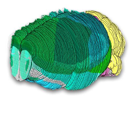
A set of articles published in Nature and Nature Methods draws a high-resolution map of the structure of and connections between the brain cells of mice. The map is based on data from a single cubic millimetre of brain and includes more than 200,000 cells, around 84,000 neurons and 524 million synaptic connections. Although this is a very small part of the mouse brain, it will help us understand how different types of cells work together.
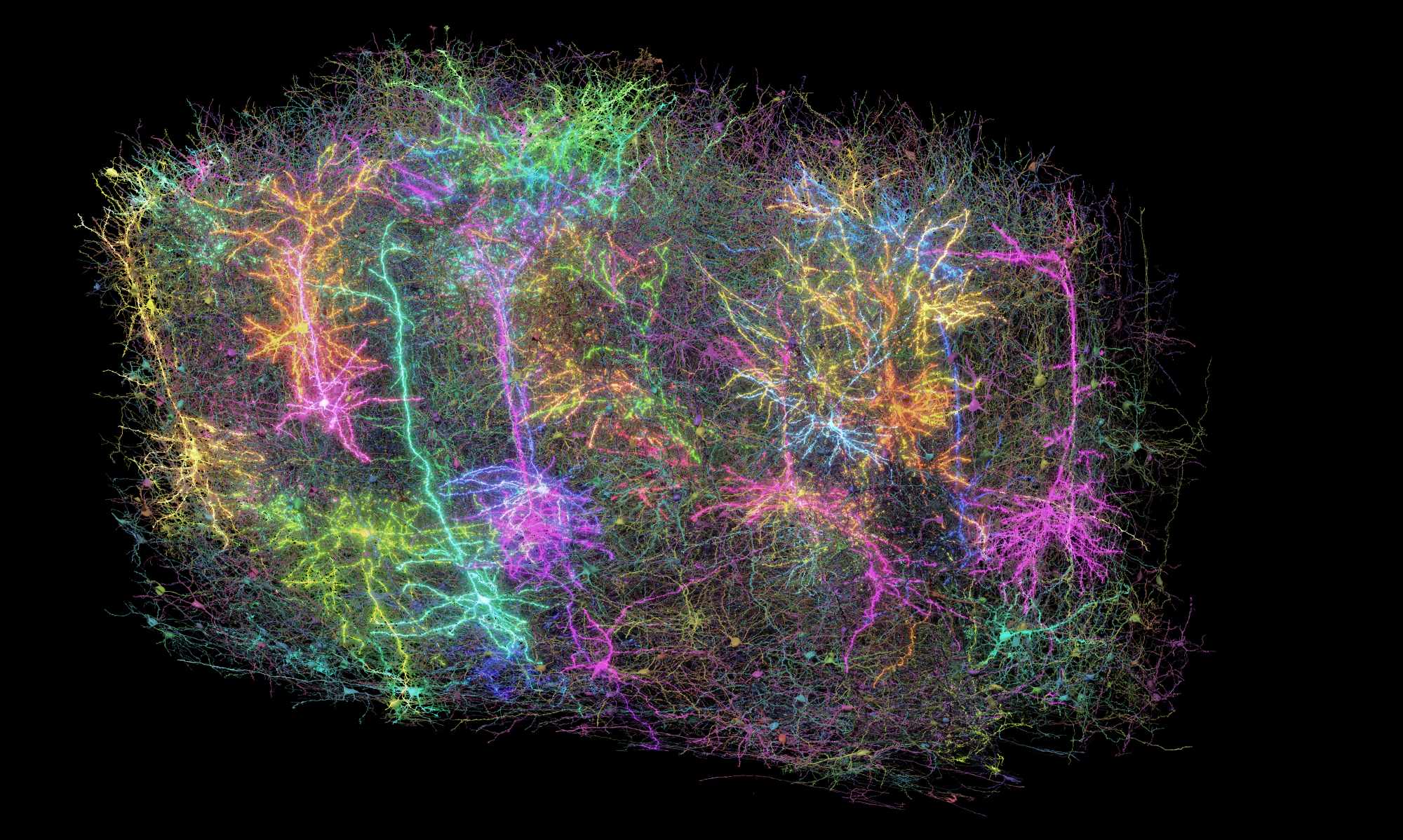
A subset of more than 1,000 of the 120,000 brain cells (neuron + glia) reconstructed in the MICRONS project. Each reconstructed neuron is a different random color. In some of the images, a subset of the neurons have been rendered as glowing in different ways to represent the fact that this dataset includes functional recordings from a subset of neurons. It is meant as a symbolic representation, but not a literal representation of what the dataset is. Credit: Allen Institute
Rafael Yuste - MICrONS EN
Rafael Yuste
Professor of Biological Sciences and Director of the Center for NeuroTechnology at Columbia University (New York), President of the NeuroRights Foundation and promoter of the BRAIN project
This batch of articles is one of the most impressive results of the BRAIN initiative [Brain Research through Advancing Innovative Neurotechnologies], which was launched by President Obama in 2013 and will last until 2030. In this case, they have used a technique called connectomics to reconstruct the circuits of the mouse cerebral cortex. It has taken almost a decade, but a consortium of laboratories has managed to use electron microscopy to map all the neuronal connections in small circuits of neurons whose activity had been measured. It is a tour de force with a wealth of results, which is like putting one of many bricks in a huge building to understand the brain.
It is worth remembering that more than a year ago another batch of impressive articles was published, mapping the cell types of the brain, which are the neurons that generate all these connections. This is another of the most impressive results to emerge from the US BRAIN project.
Now the big challenge is to bring these two strands of new science together; in other words, to understand which connections arise from which type of neuron. This will quite possibly require new techniques, such as the use of expansion optical microscopy, which allows us to do both at the same time. We now know how many types of cells there are and what the connections of all of them are like, but we need to map the connections of each type of neuron. This new form of microscopy has just been invented and I think it will go a long way.
My last comment is that these two large bodies of results, each of them worthy of a Nobel Prize in my humble opinion, have arisen not from the work of individual laboratories, but from large consortia. This is a new way of working in neuroscience and it is very similar to what happened over a decade ago in genetics with the sequencing of the human genome, and to what has been happening in physics, chemistry and astronomy for almost a century now. It is a revolution in neuroscience, not only because of the techniques or the results, but because of the way of working.
Juan Lerma - cerebro ratón conexiones EN
Juan Lerma
CSIC research professor (honorary) at the Instituto de Neurociencias de Alicante (CSIC-UMH) and member of the Royal Academy of Sciences of Spain
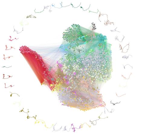
The MICrONS project provides a dataset of unprecedented scale and resolution. This dataset combines functional recordings with the high-resolution anatomical structure of several mouse visual cortical areas. Thus, more than 70,000 excitatory neurons and their responses to videos of natural scenes spanning 1 mm3 of the visual cortex were functionally recorded. This work reveals the detailed structure of approximately 60,000 excitatory neurons and 500 million synapses, representing the largest neocortical structure-function study conducted to date.
This body of work lays many of the foundations for several principles of functional organization that, although assumed, were unproven and represented gaps in our understanding of the nervous system. In fact, the connectivity principles now revealed at the structural and functional levels appear to play a fundamental role in sensory processing and learning. Furthermore, it is noteworthy that these principles are shared by both biological and artificial systems, so they represent fundamental information when designing artificial networks based on artificial intelligence. Collectively, these findings undoubtedly represent a long-awaited giant step forward, and are merely the tip of the iceberg of what is to come in understanding how the brain works. In fact, the authors also demonstrate that artificial recurrent neural networks trained on a simple classification task develop connectivity patterns that mimic the connectivity rules revealed by biological data. In short, the system's ability to process information and store it in the form of memory is determined by the connectivity of the system itself.
Furthermore, the authors have generated a basic artificial model that not only predicts neuronal activity in the visual cortex, but also the functional properties of neurons and their anatomical characteristics. In my view, these findings are of paramount importance. They will have a major impact on the field of neurocomputing and will facilitate further discoveries about the functional organization of neural circuits by allowing the exploration of previously unconsidered questions.
Both this data collection and the openly available models can be very useful for exploring which specific circuit dysfunctions could lead to functional alterations compatible with known pathologies.
María Figueres Oñate - conexiones ratones EN
María Figueres Oñate
Postdoctoral researcher at the Instituto de Investigaciones Raras Health Institute Carlos III
These multi-centre and collaborative studies are of great importance to the field, as the most significant technological advances require the participation of experts in various disciplines, capable of integrating information from multiple perspectives. Moreover, with the development of computational tools such as machine learning and artificial intelligence, the capacity for data analysis and integration has reached unprecedented levels.
However, it is crucial to remember that neurons do not act in isolation: they need a whole network of non-neuronal cells, such as glia, to establish synapses and function properly. It is therefore crucial to conduct studies that collect information in a comprehensive way, without artificially separating cells by phenotype or individual characteristics. Although specific segmentation is performed in the study, the identification of non-neuronal structures such as astrocytes, microglia and blood vessels is also achieved, which allows for a more complete view of the circuit.
This type of work, which considers the connections within a sensory system, in this case visual, including all the cellular components involved, is essential to advance our understanding of how information is processed and integrated in the brain and, ultimately, how we perceive the world around us.
The MICrONS Consortium.
- Research article
- Peer reviewed
- Animals