The most extensive gene expression map of the prefrontal cortex suggests that the maturation process is completed around age 24
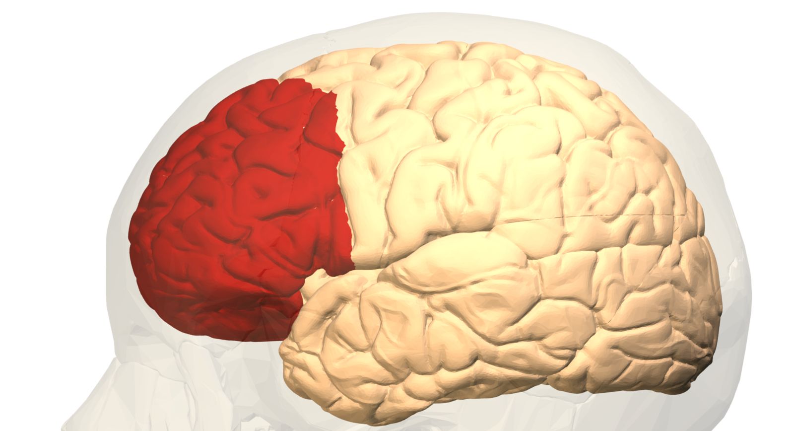
The prefrontal cortex is a brain region involved in some of the defining characteristics of human beings, such as planning, complex problem-solving, and impulse regulation. Researchers from the PsychAD consortium have created the most comprehensive map to date of gene expression in this region across the lifespan. According to the authors, the maturation process is complete by approximately age 24. The study—which will facilitate research into aspects of aging and cognitive decline—is published in Nature alongside seven other papers describing disease-related alterations and new genetic risk factors.
Lerma - Atlas corteza
Juan Lerma
CSIC research professor (honorary) at the Instituto de Neurociencias de Alicante (CSIC-UMH) and member of the Royal Academy of Sciences of Spain
This study presents a transcriptomic atlas based on single nuclei from the human dorsolateral prefrontal cortex, spanning the entire lifespan—from 0 to 97 years of age—using samples from 284 donors. This region of the cortex is involved in key cognitive functions such as problem-solving, planning, and emotional regulation, and is among the areas most susceptible to age-related decline.
The atlas reveals that gene expression in the cells of this cortex follows a non-linear trajectory throughout life. Changes are particularly pronounced during early development—including gliogenesis, synaptic remodeling, and apoptosis—followed by stabilization during adulthood. It also conclusively confirms that the age of 24 marks a turning point; beyond this age, the composition of most neuronal and glial subclasses remains stable, with few exceptions. In other words, cerebral cortex maturation extends beyond childhood and adolescence, concluding at age 24—the point marking the end of youth and the onset of adulthood. This is highly significant, as the data clarify the age up to which an individual may be considered immature and outline the potential impact of inadequate educational systems, social media exposure, and other alienating influences (such as cults, drugs, or alcohol) on brain development.
In summary, the atlas provides a foundation for understanding the cellular program transitions that shift the brain from a state of resilience to one of vulnerability. Indeed, one of the identified trajectories can be interpreted as representing neuronal resilience, as it shows reduced expression of genes associated with the risk of neurodevelopmental disorders, peaking around ages 12–13. This suggests that alterations induced prior to this critical window could have long-term functional consequences. On the other hand, defining the various turning points during development and aging may represent windows of opportunity for therapeutic intervention and guide the design of strategies to preserve brain health and reduce age-related cognitive decline.
Nieto - Atlas corteza
Marta Nieto
Head of the Cerebral Cortex Development research group at the National Center for Biotechnology (CNB-CSIC)
To reconstruct how the human brain changes over a lifetime, researchers analyzed tissue from the dorsolateral prefrontal cortex obtained post-mortem from 284 individuals, ranging in age from infancy to 97 years. From these samples, they isolated cell nuclei and used single-nucleus RNA sequencing to determine which genes were active in each cell type, creating an extraordinary atlas comprising over 1.3 million cells. The immense value of this study lies in its ability to track—separately—how each of the brain's major cell populations changes with age. Rather than viewing brain tissue aging as a single, uniform phenomenon, researchers can reconstruct the changes occurring in neurons, astrocytes, oligodendrocytes, and microglia over the course of nearly a century of human life. This description at cellular resolution is a highly valuable contribution in itself, revealing that different cell types age neither in the same way nor at the same time.
It is precisely when comparing these trajectories that the results reveal a somewhat unexpected story. During childhood and adolescence—as one might expect—neurons undergo the most significant changes as brain circuits form and mature. This is followed by a long period of relative stability. However, upon reaching old age, the brain undergoes further transformation; surprisingly, the key players this time are not the neurons, but the cells that surround and sustain them. Astrocytes and oligodendrocytes—glial cells essential for neuronal support, nutrition, and function—along with microglia (the brain's primary resident immune population), undergo profound changes in activity, upregulating programs related to cellular stress, homeostasis, and immune response.
The study also reveals another striking change. Since the researchers knew the time of death for many of the donors, they were able to reconstruct daily gene activity rhythms; they observed that the strong circadian clock synchronization seen in neurons during adulthood is largely lost after age 60, while certain glial cells and microglia acquire new rhythms linked to cellular stress.
This work thus shifts the perspective from which we view brain aging: perhaps to understand why neurons become vulnerable with age, we should look not only at the neurons themselves but also at how the cells that nourish, protect, and accompany them throughout life age.
In my view, this is a study of extraordinary quality and technical power. Beyond its immediate conclusions, its primary value lies in the vast amount of information it generates: over a million individual transcriptomic profiles, organized by cell type and spanning virtually the entire human lifespan. The study thus creates an exceptional reference resource, providing data and tools that other researchers can reuse to formulate new questions, cross-check experimental results, or explore genes, cell types, and processes associated with development and aging. As is often the case with major biological atlases, a significant portion of this work's impact will likely emerge in the coming years, as the scientific community begins to leverage information that extends far beyond the questions originally posed by the authors.
As with large-scale transcriptomic studies of human tissue, these results are fundamentally descriptive and do not demonstrate cause-and-effect relationships. The study identifies which cell types and molecular programs change with age but cannot establish whether, for instance, microglial activation or glial stress responses are a cause of neuronal vulnerability, a consequence of it, or even a protective response. Rather than a specific weakness of the study, this is a limitation inherent to this type of approach, which relies on the observation of post-mortem human tissue. Its immense value lies precisely in uncovering associations, identifying unexpected mechanisms, and generating new hypotheses that must subsequently be tested through experimental and functional studies. This does not diminish the work's value; on the contrary, the breadth and quality of the data make this atlas an excellent starting point for guiding many of the questions that research into brain aging will need to address in the coming years.
Yuste - Atlas corteza
Rafael Yuste
Professor of Biological Sciences and Director of the Center for NeuroTechnology at Columbia University (New York), President of the NeuroRights Foundation and promoter of the BRAIN project
This batch of papers is another outcome of the BRAIN Initiative launched by Obama in 2013—an initiative that continues to move forward regardless of changes in the presidency or the political climate.
This is high-quality work that employs single-cell transcriptomics to map cell types in the human prefrontal cortex. It meticulously applies methods previously established by other groups—following in the footsteps of the Allen Institute in Seattle (USA), which pioneered the use of these techniques to map cell types in both mouse and human cortices. Nevertheless, the study remains significant, as information regarding cell types is essential for understanding how neuronal circuits function.
Mapping every cell type in the brain was Cajal’s dream, and doing so in the human prefrontal cortex is particularly important, as this region is the hub for cognitive and mental information processing.
Yang et al.
- Research article
- Peer reviewed
- People